 RSS Feed
 Twitter
 6:24 PM
 Unknown
The Structure of Human Eye
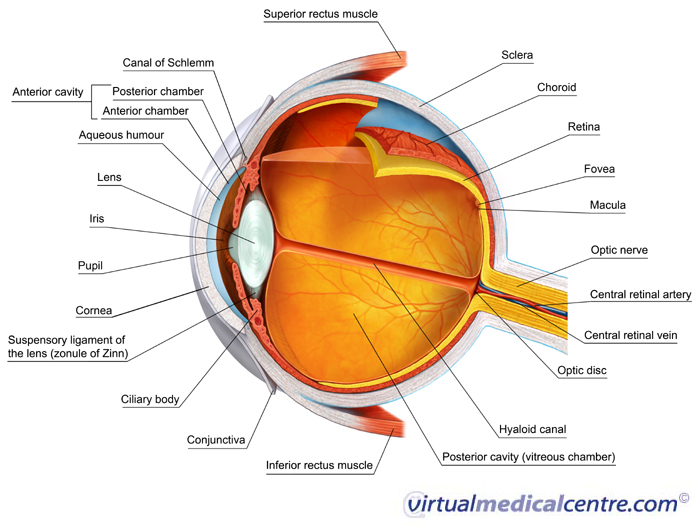
1. Ciliary body- contacts and relaxes to change the thickness of the lens. This changes the focal length of the lens.
2. Suspensory ligaments-hold the lens in place and connect it to the ciliary body.
3. Aqueous humour-helps to refract light and focus the image into retina, maintains the shape of the eye and the pressure in the eye.
4. Conjunctiva-the thin, transparent membrane that protects the cornea.
5. The pupil allows light to pass into the eye
6. Cornea-curved, transparent layer at the front of the eye. It is a continuation of the sclera. Allows light to enter the eye. Helps tofocus light onto the retina by bending the light rays passing through it.
7. Iris controls the size of the pupil and hence controls the amount of light entering the eye.
8. Eye lens-Bends and focuses light to form an image on the retina. The thickness of the lens can be altered to focus near and distant objects
9. Vitreous humour-the jelli-like material found between the lens and the retina. Helps keep the shape of the eye spherical. Helpsrefract light onto the retina.
10. Sclera pratects and shapes the eye.
11. Choroid-the capillaries supply nutrients and oxygen to the eye. The black pigments absorb light and prevent reflection of light.
12. Retina-detects light stimuli and sends nerve impulses to the brain.
13. Yellow spot (fovea)-the part of the retina most sensitive to light. Detects the images of objects formed and changes them into nerve impulses.
14. Blind spot-this point on the retina is not sensitive to light. Images falling on this spot cannot be detected because there is no receptor cells. It is the spot where the optic nerve leaves the eyeball.
15. Optic nerve-sends nerve impulses from the retina to the brain to be interpreted.
THE MECHANISM OF SIGHT

1. Light rays travel from an object and enter the eye
through the pupil.
2.
The light rays are refracted (bent) by the cornea, aqueous humour, eye lens and
vitreous humour.
3. An inverted image smaller than the actual object is
formed on retina.
If you more understand about structure and mecanism sight of eye you can see this ppt and video:
If you more understand about structure and mecanism sight of eye you can see this ppt and video:


0 komentar:
Posting Komentar